Services
Follicular Study
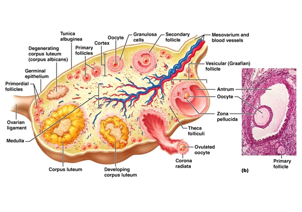
A follicular study examines the ovarian follicles using a transvaginal scan. The tissues containing eggs and the thickness of the endometrial lining are studied to determine the period that the patient is likely to ovulate.
When the eggs are mature, patients are advised to have planned relations or Intrauterine Insemination or proceed with egg collection in case of an In-Vitro Fertilization Cycle. Generally, these scans will start around day 9 of the cycle and continue till day 20 that is around 8 to 11 scans. It is a vital process for getting pregnant including through fertility treatments like IVF.
Saline Infusion Sonohysterography (SIS)
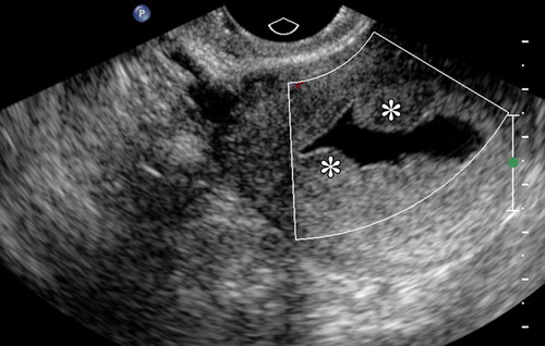
Saline infusion sonohysterography (SIS) or saline ultrasound uterine scan uses a small amount of saline (salt solution) inserted into the uterus (or womb) that allows the lining of the uterus (endometrium) to be clearly seen on an ultrasound scan.
“Ultrasound” is the term used for creating pictures or images using high-frequency soundwaves. The pictures are obtained using an ultrasound transducer.
Ultrasound transducers transmit high-frequency sound waves that are converted into electrical impulses that produce a moving image of the inside of the body on a screen.
USG (PELVIS &OBST)
USG refers to Ultrasound Sonography Test which is a diagnostic procedure used to scan the internal organs of the body by using high-frequency sound waves. USG pelvis refers to a pelvic ultrasound that is used to take pictures of the inside of the pelvis. This ultrasound is suggested to detect a particular disease or even to analyse the health of the baby in the womb of a pregnant woman. A USG pelvis is also known by several other names such as gynaecologic ultrasound, pelvic scan, pelvic sonography, transrectal ultrasound, etc.
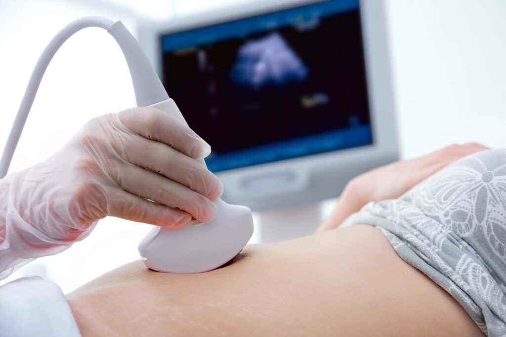
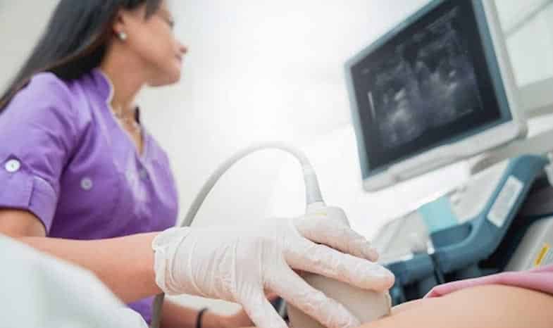
USG Pelvis produces pictures of the internal structures and organs of the area by using high-frequency sound waves. To conduct an USG Pelvis, a small probe known as transducer and gel is applied on the skin directly, to allow the high-frequency sound waves from the transducer to travel from the probe into the skin to produce the real-time images of the area, as well as the movement of the organs and blood flow in the blood vessels.
Transvaginal Sonography (TVS)
An ultrasound test uses high-frequency sound waves to create images of your internal organs. Imaging tests can identify abnormalities and help doctors diagnose conditions.
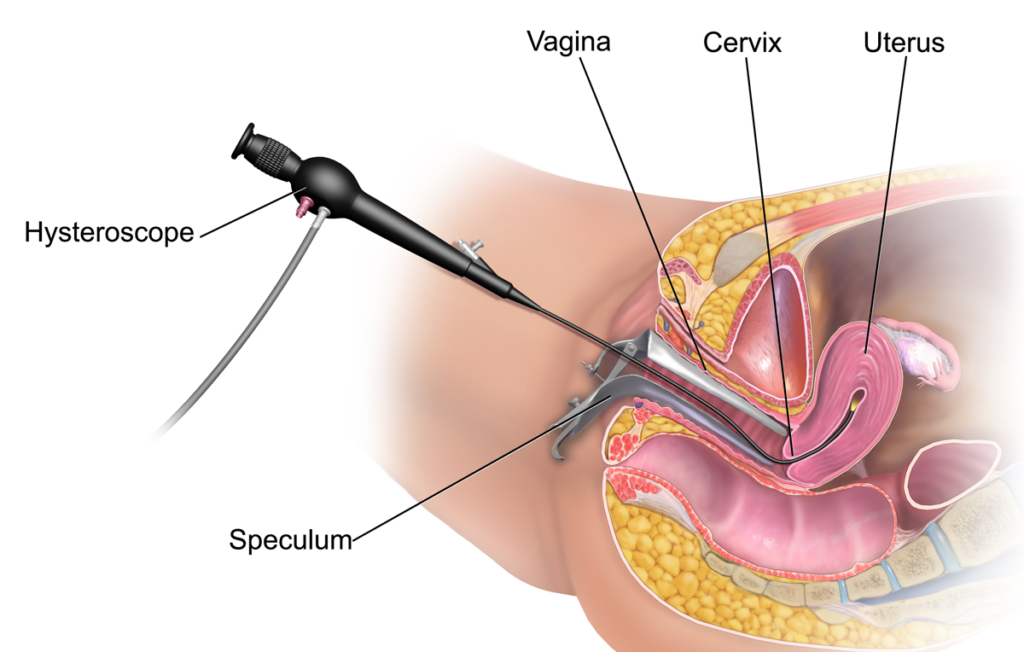
A transvaginal ultrasound, also called an endovaginal ultrasound, is a type of pelvic ultrasound used by doctors to examine female reproductive organs. This includes the uterus, fallopian tubes, ovaries, cervix, and vagina.
“Transvaginal” means “through the vagina.” This is an internal examination.
Unlike a regular abdominal or pelvic ultrasound, where the ultrasound wand (transducer) rests on the outside of the pelvis, this procedure involves your doctor or a technician inserting an ultrasound probe about 2 or 3 inches into your vaginal canal.
Intrauterine insemination (IUI)
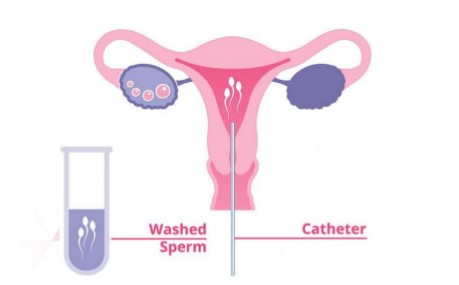
Intrauterine insemination (IUI) — a type of artificial insemination — is a procedure for treating infertility.
Sperm that have been washed and concentrated are placed directly in your uterus around the time your ovary releases one or more eggs to be fertilized.
The hoped-for outcome of intrauterine insemination is for the sperm to swim into the fallopian tube and fertilize a waiting egg, resulting in a normal pregnancy. Depending on the reasons for infertility, IUI can be coordinated with your normal cycle or with fertility medications.
Semen Analysis by Digital Workstation
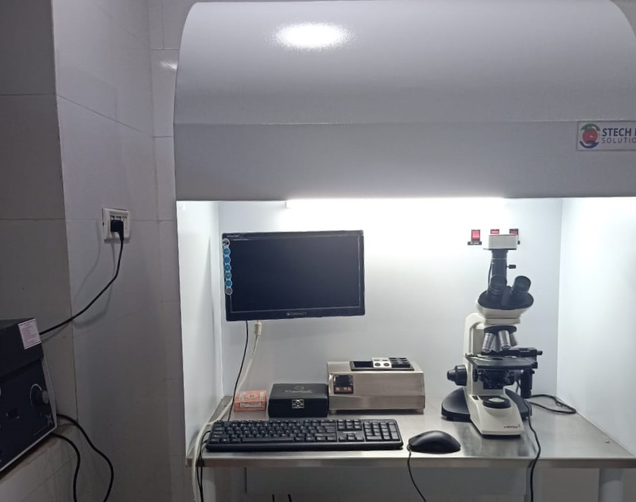
A semen analysis (plural: semen analyses), also called seminogram, or spermiogram evaluates certain characteristics of a male's semen and the sperm contained therein. It is done to help evaluate male fertility, whether for those seeking pregnancy or verifying the success of vasectomy. Depending on the measurement method, just a few characteristics may be evaluated (such as with a home kit) or many characteristics may be evaluated (generally by a diagnostic laboratory). Collection techniques and precise measurement method may influence results.
Cervical Pap Smear Screening Test
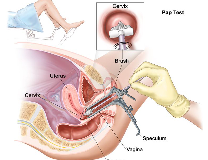
A Pap smear, also called a Pap test, is a screening procedure for cervical cancer. It tests for the presence of precancerous or cancerous cells on your cervix. The cervix is the opening of the uterus.
During the routine procedure, cells from your cervix are gently scraped away and examined for abnormal growth. The procedure is done at your doctor’s office. It may be mildly uncomfortable, but doesn’t usually cause any long-term pain.
Keep reading to learn more about who needs a Pap smear, what to expect during the procedure, how frequently you should have a Pap smear test, and more.
Colposcopy
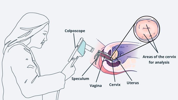
Colposcopy is the magnified visual inspection of the cervix to check for any abnormalities. In other words, your doctor or nurse is going to look at your cervix to check for any signs of problematic cells growing.
If you have an abnormal Pap test result or your HPV test came back positive, then your doctor or nurse may send you to have a colposcopy procedure. You might also have a colposcopy performed if you have experienced painful sex, unexplained vaginal bleeding or bleeding after sex to look for possible explanations of these symptoms.
Laparoscopy
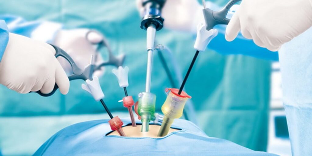
Laparoscopy, also known as diagnostic laparoscopy, is a surgical diagnostic procedure used to examine the organs inside the abdomen. It’s a low-risk, minimally invasive procedure that requires only small incisions.
Laparoscopy uses an instrument called a laparoscope to look at the abdominal organs. A laparoscope is a long, thin tube with a high-intensity light and a high-resolution camera at the front. The instrument is inserted through an incision in the abdominal wall. As it moves along, the camera sends images to a video monitor.
Laparoscopy allows your doctor to see inside your body in real time, without open surgery. Your doctor also can obtain biopsy samples during this procedure.
